

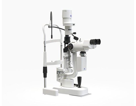
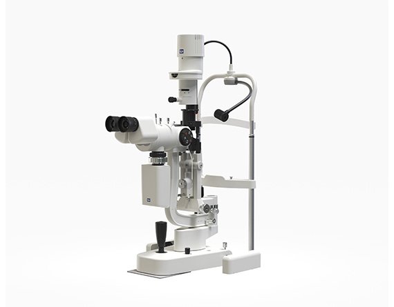
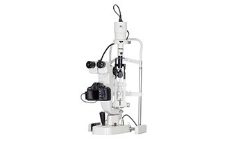
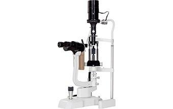
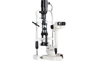
YZ5T Slit Lamp Imaging System
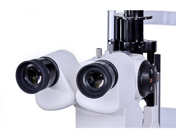
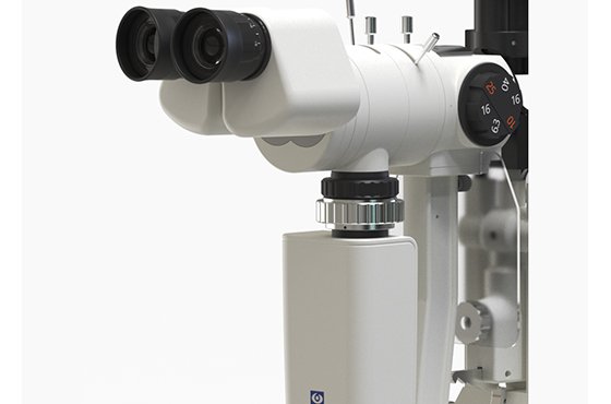
- Galilean stereoscopic microscope
- German optical lens for clear and sharp image
- Five drum rotating manifications
- Multi-layer coatings for anti-reflection, moisture-proof, and mold-proof
- Halogen/LED light source for option
New and Star Slit Lamp by 66 Vision
YZ5T slit lamp imaging system is an image processing and patient data managing system developed by 66 Vision. It is designed to meet the needs of eye care professionals worldwide. It is used to analyze and process the photos and videos captured by the digital slit lamp.
Specifications
| Microscope type | Galilean Stereoscopic Microscope |
| Objective magnification | Five-step drum magnification |
| Eyepiece | 12.5X |
| Magnification(Field of view) | 6.3X(𝞍33mm),10X(𝞍22.5mm),16X(𝞍14mm),25X(𝞍8.8mm),40X(𝞍5.5mm) |
| Pupil distance | 52mm~80mm |
| Diopter adjustment | -8D~+8D |
| Slit width | 0mm~14mm adjustable(slit is round when the slit width is 14mm) |
| Slit height | 1mm~14mm adjustable |
| Diameter of light spot | 𝞍14mm, 𝞍10mm, 𝞍5mm, 𝞍2mm, 𝞍1mm, 𝞍0.2mm |
| Slit angle | 0°~180°adjustable |
| Slit inclination | 5°, 10°, 15°, 20° |
| Filters | Heat absorption, Grey, Redfree, Cobalt blue, Yellow Filter Module |
| Illumination source | 12V30W halogen bulb/LED |
| Collecting medium | CCD |
| Illumination type of taking photo | Background illumination, space fixation |
| Power supply | AC110V/220V±10%, 50Hz/60Hz |
| Optional accessories | Appanation tonometer, Measuring eyepiece, Trihedron lens, Gonioscope, Funduscope, Aspheric lens |
| Dynamic display | Computer screen real-time dynamic display |
| Packing volume | 670mm*620mm*490mm |
| Total weight | 23kg(table and computer not included) |
| Processing function | Length, Angle, Area, Measuring, Arrowhead, Word adding, Image processing |
| Table (optional) | YT2GB |
Global Photography Technology
Sony Global shutter CMOS, applied in the YZ5T slit lamp, with integrated depth of field, to capture dynamic image, revealing detailed lesions.
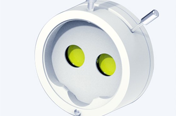
Built-in Yellow Filter
A newly added yellow filter highlighting corneal fluorescein allows strong contrast of sodium-stained images for early detection of lesions.
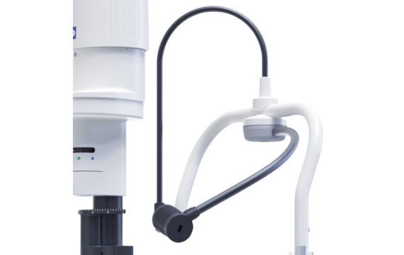
Space Fixation and Background Lighting
Independent target fixation and background light design for free space adjustment to meet multi-angle observation needs
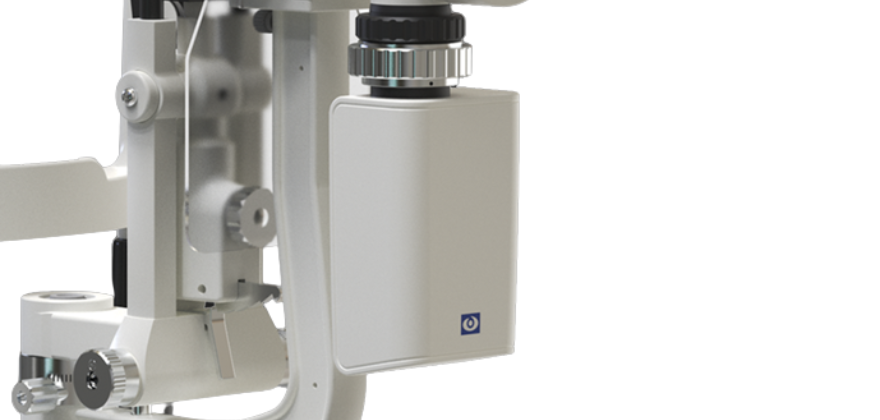
Outstanding Medical CCD Camera
Our YZ5T digital slit lamp uses medical CCD image sensors to obtain images. Medical CCD cameras have good color reproduction capabilities, high image quality and signal-to-noise ratio, and can capture very clear and delicate images. In addition, CCD cameras have high sensitivity and can capture tiny signals, which is of great significance for the diagnosis of tiny lesions. Moreover, CCD can image in real time, so doctors can immediately observe the status of the lesion, thereby shortening the diagnosis time.
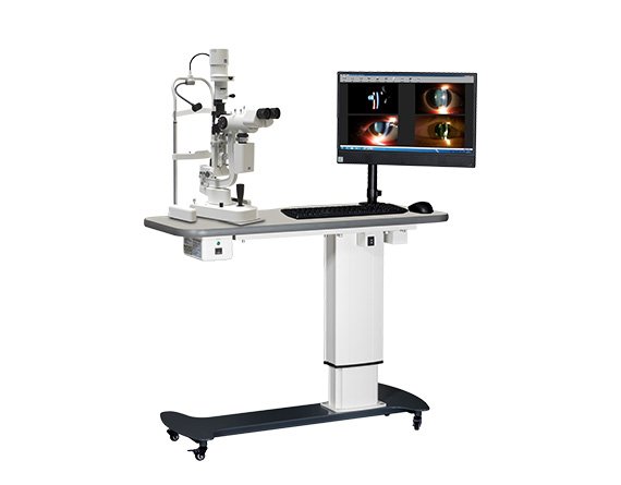
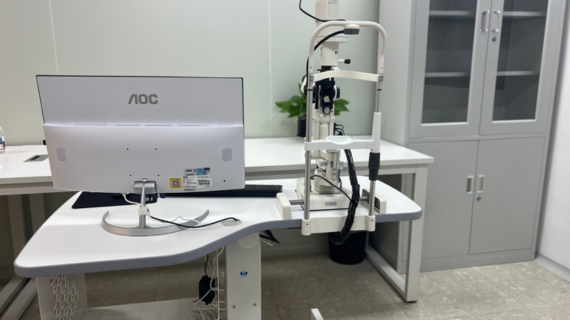
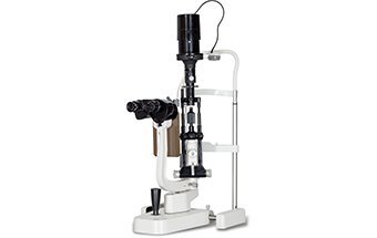
YZ5T Slit Lamp Components and Packing
YZ5T is a complete set of slit lamp microscope, including slit lamp, CCD camera, computer, software and motorised table. It is packed in three cartons. Each carton has two layers to protect the slit lamp during tranportation.
Eye Image Gallery
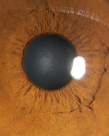
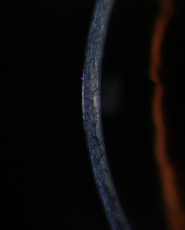
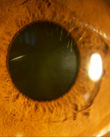
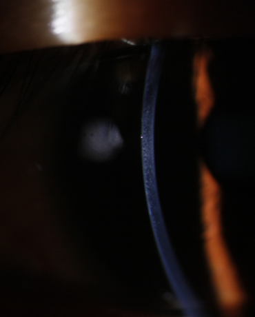
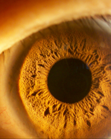
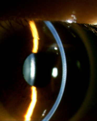

We use medical CCD camera to capture images. The CCD cameras are often used in operation microscopes, they can also take videos.
We have a full stock of YZ5T digital imaging slit lamp. So we can ship out as soon as we receive the payment.
Yes, we offer the software for free.
The yellow filter of the slit lamp is mainly used to enhance the observation of irregular staining on the corneal surface. Especially in the sodium fluorescein staining examination, it can significantly improve the contrast of the lesion area by using it in combination with cobalt blue light.